PLANTAR HEEL PAIN: AN EVIDENCE-BASED APPROACH TO DIAGNOSIS AND TREATMENT

You might have come across patients complaining about pain on the plantar surface of the heel, well, plantar heel pain is a highly prevalent condition in both non-athletic and athletic populations. Despite its prevalence, little is known about the pathology, risk factors, prognosis, and the best possible treatment.
It can be a very tricky condition to treat and might take quite some time in order for us to give the best possible treatment to our patients.
PATIENT’S COMPLAINT
The patients with plantar heel pain usually complain of having the “first step pain”. It can be the first step after getting out of bed or the first step after the patient have been sitting down for a long period of time. The Patient usually complain of pain while standing up and walking around.
A couple of things you should keep in mind about plantar heel pain that will assist you in the differential diagnosis are:
- The plantar heel pain usually improves with ambulation. While heel pains due to other conditions like fat pad atrophy, fat pad contusion, or plantar fascia rupture, the pain gets worse with ambulation.
- In the case of plantar heel pain, the onset of pain is usually insidious, while in most other cases of heel pain, it is usually acute. Both of these things will help you in the differential diagnosis of plantar heel pain.
In the case of plantar heel pain, the pain point is usually located at the site where the plantar fascia inserts into the calcaneus. Plantar heel pain is quite prevalent in the general population, especially in running athletes. It is also the cause of many other running injuries. In fact, 8% of all running injuries are caused by plantar heel pain.
Evidence suggests that people with plantar heel pain exhibit higher level of depression (1). It is also demonstrated that there is an association between plantar heel pain, kinesiophobia, pain catastrophizing, and pain and function (2).
Based on this evidence, addressing the adjoining psychological factors using the biopsychosocial model becomes an integral part of treatment for plantar heel pain.
DIAGNOSIS
The patient’s history and palpation are sufficient to establish the diagnosis of plantar heel pain. Ultrasound can also be considered to establish the diagnosis.
HISTORY AND PALPATION
The patient’s history can be the initial guide toward establishing the diagnosis of plantar heel pain. Palpation of the plantar heel is the next step in the examination.
Pain with palpation of the proximal insertion of the plantar fascia is one of the signs suggesting plantar heel pain. The pain needs not to be localized. Pain can extend well beyond the plantar heel. If this is the case, you might consider ultrasound imaging of the plantar heel to establish the diagnosis.
The recent evidence suggests that patients with plantar heel pain are 105 times more likely to have a plantar fascia thickness ≥4.0 mm compared to pain-free controls (3). So, ultrasound might help establish the diagnosis where the presenting symptoms are somewhat non-specific.
RECENT CLINICAL GUIDELINES
Recent clinical guidelines include a checklist to rule out other causes of heel pain and confirm the diagnosis of plantar heel pain. The checklist include:
- Positive windlass test
- Negative tarsal tunnel test (Tinel’s sign)
- Limited ankle ROM
- Abnormal foot posture index
- High BMI in non-athletic population
All of these need not to be fulfilled to establish a diagnosis of plantar heel pain. The most important are negative tarsal tunnel test and positive windlass test.
TARSAL TUNNEL SYNDROME
Two tests most commonly used to rule out tarsal tunnel syndrome are:
- Triple compression test
- Tinel’s sign
Triple compression test
To perform a triple compression test, the patient is supine lying. The examiner performs a maximal plantarflexion of the ankle along with maximal inversion of the foot and heel. The examiner applies sustained pressure over the tibial nerve posterior to the medial malleolus. The test is positive for tarsal tunnel syndrome if the patient experiences numbness, paraesthesia, or pain along the distribution of the tibial nerve.
Tinel’s sign
It is easy to perform. Lightly tab along the tibial nerve posterior to the medial malleolus. The test is positive if the patient experiences numbness, paraesthesia, or pain along the tibial nerve.
Tarsal tunnel syndrome needs to be ruled out in order to confirm the diagnosis of plantar heel pain.
POSITIVE WINDLASS TEST
Windlass test performed in standing position with the head of metatarsals on the edge of chair or stool. The patient puts weight on the leg. The test is positive if the dorsiflexion of the big toe produces pain at the insertion of the plantar fascia or the head of the first metatarsal.
A positive windlass test and a negative tarsal tunnel test are needed to confirm the diagnosis of plantar heel pain. The rest of the checklist may or may not be present.
HEEL SPUR
Most of you might think that the heel spur should be the main culprit for plantar heel pain. Well, this is not true. Only 2-3% of people with plantar heel pain have a heel spur confirmed by X-ray or Ultrasound. A heel spur is a calcification on the calcaneus. It might be a common finding in people with no heel pain, and they might never develop one.
PLANTAR HEEL PAIN OR PLANTAR FASCIITIS
Previous studies suggested that there were no signs of inflammation in the plantar fascia and that only degenerative changes were present in the case of plantar heel pain. These studies suggested not using the term plantar fasciitis but using plantar heel pain (4).
But recent studies have shown that tendinopathy involves chronic inflammation and argued that the term, plantar fasciitis, can be used alternatively for plantar heel pain (5).
DIFFERENTIAL DIAGNOSES
As a physiotherapist, you must rule out other causes of heel pain. The possible differential diagnoses for heel pain are:
FAT PAD ATROPHY
It is often seen in older adults and in contrast to plantar heel pain, fat pad atrophy gets worse with ambulation.
FAT PAD CONTUSION
Fat pad contusion has an acute onset while plantar heel pain has an insidious onset and fat pad contusion does not get better with ambulation.
SPONDYLOARTHROPATHIES
In case a patient has spondyloarthropathy, the patient will also complaint of low back pain (LBP), and the patient usually presents with bilateral symptoms, and bilateral joint complaints.
BAXTER’S NEUROPATHY
In case of Baxter’s neuropathy, there will be positive tarsal tunnel test along with pain on palpation of medial aspect of plantar heel.
PLANTAR FIBROMA
In case of plantar fibroma, there will be a palpable lump in the plantar fascia.
CALCANEAL STRESS FRACTURE
Calcaneal stress fracture has symptoms quite similar to fat pad contusion, like acute onset, pain not getting better with ambulation. But resting pain and positive squeeze test of heel are unique to calcaneal stress fracture.
PLANTAR FASCIA RUPTURE
Plantar fascia rupture has an acute onset. The pain is more distal than plantar heel pain. The pain does not improve with ambulation.
PROGNOSIS
Having an idea about the prognosis of a specific disease or dysfunction is extremely necessary while practicing in clinical settings. Some patients might ask, will it ever go away? Or some patients might want to know how long it will take to be completely free of pain, dysfunction, or disease. In these circumstances, you must have an evidence-based idea of the prognosis of the dysfunction or disease.
If a patient does not ask about the prognosis, you should ask them about their expectations regarding their recovery. It will give you an idea about the patient’s hopes and goals regarding their recovery so you can choose and plan the treatment accordingly.
In the case of plantar heel pain, tell the patient that it might take a couple of months if treated, and for some, it might take even longer. Evidence shows that 40% of the patients with plantar heel pain experience symptoms two years after treatment (6). In more severe cases, 50% will have symptoms ten years after treatment (7).
Below are certain factors that indicate a poor prognosis of patients with plantar heel pain:
- Symptom duration > 7 months
- Gender: Female
- Age < 40 years
- Bilateral pain
These factors indicate a poor prognosis, and it might take several years for the plantar heel pain to subside in these patients.
Certain factors that we think as important determinants of the prognosis of plantar heel pain, like High BMI, Plantar fascia thickness, and the presence of heel spur, have little or even no role in establishing the prognosis of plantar heel pain.
TYPICAL PATIENT
The typical patient presenting with plantar heel pain usually ranges from an athlete with a large training volume to a sedentary middle-aged person with a high BMI and everyone in between them.

RISK FACTORS FOR PLANTAR HEEL PAIN
Different risk factors for developing plantar heel pain have been identified in different populations. It usually includes Non-athletic and athletic populations.
RISK FACTORS IN THE NON-ATHLETIC POPULATION
- High BMI; People with or at risk of developing plantar heel pain should be followed over an extended period of time to see whether BMI increases first, leading to plantar heel pain, or the pain develops first, leading to less mobility resulting in a high BMI.
- Flat foot in standing
- A plantar fascia thickening
- Heel pad thickening
- Heel spur
- Baxter’s neuropathy
RISK FACTORS IN ATHLETIC POPULATION/ RUNNERS
- Genu Varum
- Use of running shoes with spikes
- Cavus arch posture
- More days running per week
- More years as an active runner
- More kilometers running per week
MECHANISM OF INJURY IN PLANTAR HEEL PAIN
Plantar heel pain is usually a type of overuse injury. It occurs when the load put on the body is greater than the capacity of the body to bear that load. There are two ways to avoid the risk of overuse injury:
- First, we can reduce the load and bring it to the level of the capacity of the body.
- Second, we can build the body's capacity and gradually increase the load as the capacity increases.
TREATMENT
As a physiotherapist, we have numerous treatment options for patients with plantar heel pain. The choice of treatment depends upon the patient. The stepwise approach should be followed in the management of plantar heel pain.
PATIENT EDUCATION
Patient education about the risk factors and mechanism of injury is extremely important in the management of plantar heel pain. If the patient is overweight, start with advice to lose weight. It might not be an easy task for the patient. The physiotherapist should explain to the patient that by decreasing weight, we are lessening the risk of developing plantar heel pain.
If the occupation of the patient involves hours of standing, advise them to modify their workstation and reduce the standing hours to reduce the strain on the plantar fascia and reduce the risk of plantar heel pain.
Advise the patient on decreasing the everyday load initially and explain to them that they can build up the capacity gradually over time and increase the load accordingly.
In the case of runners, reduce the training volume. Runners usually ask whether they should run or walk. The most appropriate answer to this question is, ”You can run as long as it does not exacerbate your symptoms. Preferably you should walk and slowly rebuild the training volume.”
An athlete should maintain or increase VO2 max by cycling, swimming, or rowing if they do not exacerbate symptoms. Advise the patient to consider the period of less running as an investment to finally becoming pain-free.
CHOICE OF TREATMENT BETWEEN FOOT ORTHOSIS AND CORTICOSTEROID INJECTION
FOOT ORTHOSIS
Foot orthosis are treatment of choice in patients with:
- Greater ankle dorsiflexion
- Lower BMI
- Lower fear-avoidance beliefs
CORTICOSTEROIDS INJECTION
Corticosteroid injection can be considered in patients with:
- Fewer weight-bearing hours
- Less baseline pain
LESS INVASIVE TREATMENTS
HEAVY SLOW RESISTANCE TRAINING
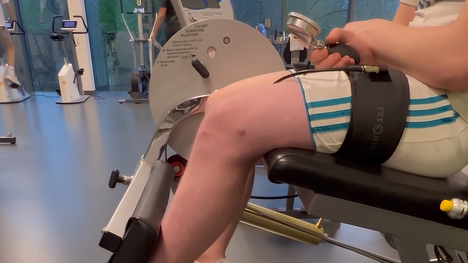
Heavy slow resistance training is used to treat tendinopathies. It produces the windlass effect, according to which strain is applied to the tendons in order for them to adapt. Stain should be high and has to surpass the strain of everyday activities.
Heavy slow resistance training usually involve unilateral heel raise with dorsal flexion of toes. Raise the heel while standing just with the forefoot on the step and then use a rolled up towel underneath the toes to dorsiflex them maximally and then do a very slow heel raise.
Raise the heel slowly and hold this for 2 seconds and then lower their heel for 3 second. It is the high load slow training.
We start by having the patient perform the heel raise with both feet and if they are able to do 15-20 repetitions, we will ask them to perform the heel raise unilaterally on the painful side.

If the resistance seems low, you can add more resistance with the help of a bag filled with weights. Heavy slow resistance training is usually considered to be painful, but this pain is different from plantar heel pain. The pain due to heavy slow resistance training does not cause any harm and should not be a barrier to performing the exercise. Patients should not skip the exercise just because it is painful.
There are usually two protocols followed for heavy slow resistance exercises:
- Pre-determined protocol
- Self-dosed protocol
Pre-determined protocol
The pre-determined protocol includes pre-set criteria for contraction time, repetitions, and sets for exercise.

Self-dosed protocol
The self-dosed protocol includes using weight as heavy as possible but no heavier than a weight that can be lifted 8 times and performing as many sets as possible.

Effect of a pre-determined protocol for heavy slow resistance training is somewhat equal to the self-dosed protocol.
It is found that with a self-dosed approach, people are more likely to comply with the exercise. In the self-dosed approach, 42 sessions over a period of 3 months are usually needed to bring a significant change in the patient’s condition.
STRETCHING
While following a stepwise approach, plantar fascia stretch is the next resort. Take the toes, straighten them, and then dorsiflex them as much as you can and feel the stretch with the other hand.
The stretching is usually done in the sitting position. The protocol usually followed for stretching plantar fascia is:
- Three times daily
- Ten repetitions with ten seconds each
Plantar fascia stretching appears to be superior to Achilles tendon stretching.
FOOT ORTHOSES, HEEL CUPS, AND INSOLES
Foot orthoses are usually better than placebo for 0-4 weeks but no longer than that. Prefabricated orthosis and foot insoles are as good as customized ones. The orthoses on their own might not show significant improvements, but when you combine them with stretching, dramatic results can be seen.
The best orthosis or insole for plantar heel pain can only be found after trying different types and then choosing one that gives immediate pain relief or comfort.
TAPING
Taping is sometimes used if the plantar heel pain is associated with flatfoot. Taping can also be used to support the arches of the foot. Plantar heel pain is usually associated with flatfoot in standing.
The taping is just a temporary solution, and strengthening of flexible flatfoot and correction of structural flatfoot might be necessary to treat plantar heel pain permanently.
ELECTRO-PHYSICAL AGENTS
Electro-physical agents like ultrasound and interferential therapy are sometimes utilized to treat plantar heel pain. Their effectiveness can be exponentially increased by combining them with other non-invasive treatment options.
EXTRACORPOREAL SHOCKWAVE THERAPY (ECSWT)
Extracorporeal shockwave therapy is being used to treat plantar heel pain. It is used to treat tendinopathies but usually gives better results in plantar heel pain than in other conditions.
A great benefit of ECSWT is both the short and long-relief in case of plantar heel pain. When the effectiveness of ECSWT and corticosteroid injection is compared for the treatment of plantar heel pain, they have shown similar outcomes. According to a meta-analysis, ECSWT works better when the patient is not anaesthetized (When they can feel the pain during treatment) compared to when the patient is anaesthetized (despite a similar dose). It suggests that ECSWT can have a better outcome in patients with plantar heel pain if the patient receives treatments when not anaesthetized.
Focused extracorporeal shockwave therapy has a slight advantage over radial extracorporeal shockwave therapy. The main problems with ECSWT treatment are its high cost and pain during treatment.
INVASIVE TREATMENT APPROACHES
INJECTION
Three types of injections most commonly used for plantar heel pain are:
- Corticosteroid
- Botulinum toxin A
- Platelet-rich plasma
Of these three, corticosteroid injection is mostly used. Corticosteroid injection is good for the short-term solution of plantar heel pain for about 4-6 weeks. It is a more invasive treatment approach along with its risks. But with the advent of ultrasound-guided corticosteroid injections, the risk of plantar fascia rupture is negligible. Ultrasound-guided corticosteroid injection is more safe and effective than palpation-guided injection.
SURGERY
Surgery is our last resort toward the treatment of plantar heel pain. If all the non-invasive treatment options and injections fail to relieve plantar heel pain, following surgical approaches might be used to treat plantar heel pain:
- Endoscopic fascial release
- Topaz Coblation
- Gastrocnemius recession
APPROACHES TO MANAGEMENT OF PLANTAR HEEL PAIN
The two most commonly used approaches to treat plantar heel pain are:
- Core Approach
- Progressive treatment approach
CORE APPROACH
The core approach has the strongest evidence for the treatment of plantar heel pain (8). According to the core approach, patient education, plantar fascia stretch, and taping are essential for every patient with plantar heel pain. After the individual assessment, you decide whether the patient needs load management advice, pain education, footwear recommendations, or others, depending upon your assessment and evaluation.

PROGRESSIVE TREATMENT APPROACH
The progressive treatment approach follows a stepwise approach from the least or non-invasive treatment to a more invasive and aggressive treatment approach.
- In a progressive treatment approach, we start with good advice, insoles, or tape, if an athlete has the urge to participate in a specific activity. We can also consider fascia stretching if the patient wants to be more active in the treatment.
- If this doesn’t work, we move on to heavy slow resistance training.
- If heavy slow resistance training doesn’t work, our next step should be extracorporeal shockwave therapy or corticosteroid injection.
- As always, if nothing else works, surgery should be considered to treat plantar heel pain.
Keep the exercises simple so the patient can understand and perform them easily.
CONCLUSION
Let’s conclude our discussion with the following points:
- There is no superiority of one treatment over another, but the treatments were better than the placebo. Using a combination of different treatment approaches is more effective than a single treatment. For example, combining corticosteroid injection with heavy slow resistance training can provide the patient with both short-term and long-term relief.
- Self-dosing in heavy slow resistance training is an alternative to the pre-determined dosing approach.
- Corticosteroid injection and extracorporeal shockwave therapy are more likely to be effective, especially in the short term.
- A novel treatment approach called percutaneous needle electrolysis (PNE) might be another treatment option for the treatment of plantar heel pain.
- Manual soft tissue treatments
- Manual soft tissue treatments can provide short-term relief. None of the treatments have superiority over the others.
- The research regarding surgery for plantar heel pain lacks randomized control trials (RCTs), and the previous research is based on case studies and comparison groups. Surgery is the last resort and might work for some but not for others. Surgery carries more adverse effects and risks than exercise.
REFERENCES
- Cotchett M, Munteanu SE, Landorf KB. Depression, Anxiety, and Stress in People With and Without Plantar Heel Pain. Foot Ankle Int. 2016;37(8):816-21.
- Cotchett M, Lennecke A, Medica VG, Whittaker GA, Bonanno DR. The association between pain catastrophising and kinesiophobia with pain and function in people with plantar heel pain. The Foot. 2017;32:8-14.
- McMillan AM, Landorf KB, Barrett JT, Menz HB, Bird AR. Diagnostic imaging for chronic plantar heel pain: a systematic review and meta-analysis. Journal of Foot and Ankle Research. 2009;2(1):32.
- Riel H, Cotchett M, Delahunt E, Rathleff MS, Vicenzino B, Weir A, et al. Is ‘plantar heel pain’ a more appropriate term than ‘plantar fasciitis’? Time to move on. British Journal of Sports Medicine. 2017;51(22):1576-7.
- Dakin SG, Newton J, Martinez FO, Hedley R, Gwilym S, Jones N, et al. Chronic inflammation is a feature of Achilles tendinopathy and rupture. British Journal of Sports Medicine. 2018;52(6):359-67.
- Caratun R, Rutkowski NA, Finestone HM. Stubborn heel pain: Treatment of plantar fasciitis using high-load strength training. Can Fam Physician. 2018;64(1):44-6.
- Hansen L, Krogh TP, Ellingsen T, Bolvig L, Fredberg U. Long-Term Prognosis of Plantar Fasciitis: A 5- to 15-Year Follow-up Study of 174 Patients With Ultrasound Examination. Orthop J Sports Med. 2018;6(3):2325967118757983.
- Morrissey D, Cotchett M, J'Bari AS, Prior T, Griffiths IB, Rathleff MS, et al. Management of plantar heel pain: a best practice guide informed by a systematic review, expert clinical reasoning and patient values. British Journal of Sports Medicine. 2021;55(19):1106-18.