ILIOTIBIAL BAND SYNDROME: THE PATHOPHYSIOLOGY, CLINICAL ASSESSMENT, DIAGNOSIS, AND TREATMENT STRATEGY

Iliotibial Band Syndrome (ITBS) is a common presenting complaint, especially in runners.
The patient presents with pain on the lateral aspect of the knee joint, where the iliotibial band inserts into Gerdy’s tubercle. The incidence and frequency of Iliotibial band syndrome (ITBS) vary widely among the population and sports.
The iliotibial band usually affects 12-52% of habitual runners and accounts for 1.6-52% of all knee injuries (1). We are more likely to see ITBS in sports like running, hockey, football, rowing, and cycling. Here we will further discuss the anatomy of the iliotibial band (ITB), the examination and assessment of individuals with iliotibial band syndrome, and diagnosis and treatment in light of recent evidence.
ANATOMY OF THE ILIOTIBIAL BAND (ITB)
Kaplan, in 1958, described it as connecting the ilia to the tibia, fusing with tensor fascia latae and gluteus maximus forming what he called the deltoid of the hip. It inserts distally into the tubercle of Gerdy, as stated by Kaplan. It is made up of avascular connective tissue, type-1 collagen, and elastin (2).
 Reproduced from Flato R, et al, 2017.
As the iliotibial band is mainly fascial tissue, it demonstrates the ability to adapt to and transfer mechanical stresses. These stresses usually involve repetitive forces at the knee joint as the individual walks or runs. For example, a runner usually takes 150 steps per minute, exposing ITB to repetitive forces.
The iliotibial band (ITB) is highly innervated and transfers nociceptive and other kinesthetic sensations and tension.
It is difficult to stretch ITB to a significant length because it is fascial tissue and requires a lot of force to produce any deformation. So regular stretches might not provide enough ITB stretch to lengthen the tissue to a significant length (3). Chaudhry et al, 2008 reported that it would take over 200kg of force to stretch the ITB by 1% (4).
UNDERSTANDING THE PATHOPHYSIOLOGY OF ILIOTIBIAL BAND SYNDROME (ITBS)
There is usually a misconception that the lateral knee pain in ITBS is caused by the friction that occurs between the iliotibial band and the lateral epicondyle of the femur, irritating the bursae immediately under the iliotibial band at the site of its insertion over Gerdy’s tubercle.
This belief was challenged by Nemeth and Sanders in 1996. They examined eight cadaveric knees and found no bursae underneath the iliotibial band at its insertion site. They also deduced the same results from the total knee replacements they performed on their patients.
Their findings revealed that the tissue under the ITB consists of synovium that is merely a lateral extension and invagination of the knee joint capsule and is not a separate bursa (5).
Recent studies conclude that a highly innervated adipose tissue resides under the iliotibial band at the site of its insertion that gets compressed during knee flexion, more precisely at 30ᵒ of knee flexion, which we call a compression zone (6). This evidence supports our understanding of why most runners feel pain in the early to mid-stance phase of the gait cycle.
 Reproduced from Sports injury bulletin by Alicia Filley.
Hip abductor strength is usually reduced in patients with ITBS. This weakness can be a contributing factor to lateral knee pain. It can also explain why some knees experience ITB pain but others do not.
A study examined 24 long-distance runners with ITBS and found reduced hip abductor strength in these runners. It is still not clear whether the reduced hip abductor strength is present before the onset of ITB pain or is a result of ITB pain. The study incorporated a hip strengthening program for six weeks and the resultant increase in hip strength made 22 out of 24 runners to return to pain-free running. One significant point of consideration is that they treated them with NSAIDs and made them pain-free before incorporating a hip strengthening program (7).
The study also showed that individuals with ITB pain in the injured limb can also have reduced strength in the non-injured limb compared with healthy individuals. The abductor strengthening program dramatically increased the abductor strength of injured and non-injured limbs and rendered the patient pain-free (7).
 Reproduced from Fredericson et al, 2000.
 Reproduced from Fredericson et al, 2000.
A prospective study by Noehren B, Davis I, and Hamill J. concluded that ITBS appears to be associated with increased hip adduction and medial rotation. These combined movements can increase the iliotibial band strain, causing it to compress against the lateral femoral epicondyle causing ITBS. These findings make it rational to consider these secondary movements as a potential cause of iliotibial band syndrome and address these movements while planning the interventions for ITBS (8).
In 2007, A study by Hamill J, Miller R, Noehren B, and Davis I. found that the major factor in the development of iliotibial band syndrome (ITBS) is the strain rate. They suggested the strain rate rather than the magnitude of strain as the causative factor for developing ITBS (9).
They also found out that the runners with iliotibial band syndrome exhibit greater strain in their iliotibial bands throughout the critical phase of gait (20-30ᵒ of knee flexion) compared with the control group.
 Reproduced from Hamill J, Miller R, Noehren B, Davis I., 2008.
CLINICAL ASSESSMENT OF INDIVIDUALS PRESENTING WITH ILIOTIBIAL BAND SYNDROME
SUBJECTIVE ASSESSMENT
Subjective assessment can be of principal importance in the individuals presenting with ITBS. The most common findings in the subjective assessment of individuals presenting with ITBS are as follows:
- A sudden increase in training volume (the most common finding), particularly an increase in hill running mainly downhill or a single increase in a long run. During downhill running, the knee is mostly in a flexed position, putting more strain on the iliotibial band and compressing the adipose tissue under the ITB, causing lateral knee pain
- The individual will complain of focal lateral knee pain following a period of running
- Listen to the individual to exclude the potential differential diagnoses like Patellofemoral pain syndrome (PFPS), Lateral collateral ligament sprain, osteoarthritis of the lateral tibiofemoral joint, and meniscal pathology.
- If there is any trauma, ask for the mechanism of injury. It can disclose valuable information that can lead to an accurate diagnosis. Because in ITBS, we are looking for more of a non-traumatic gradual onset of symptoms.
OBJECTIVE ASSESSMENT
The objective assessment for ITBS might include some special tests, assessment of the hip musculature strength, and gait analysis. Following is the updated and evidence-based objective assessment to consider while assessing the individuals presenting with lateral knee pain.
*We recommend rotating your phone to watch the videos*
Nobel’s Compression test
General knee assessment
Dynamic assessment of hip and quadriceps muscles
Assess the provocative activity with video analysis
Assessing the provocative activity with video analysis can be remarkable in finding any biomechanical abnormality, strength deficit, contralateral pelvic drop, or any other abnormality leading to increased ITB strain. Doing a three-dimensional analysis using slow-motion video can be of great help in this regard.
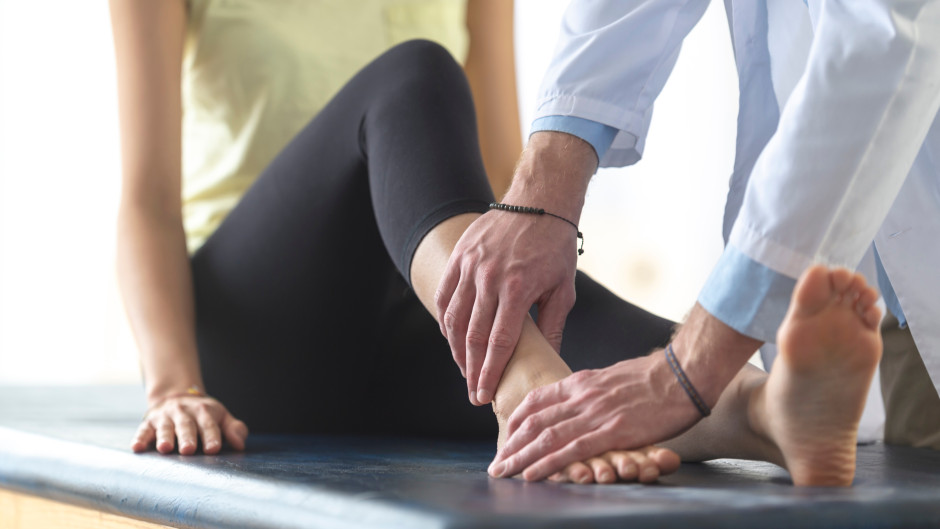
Test the strength of hip abductors and external rotators using hand-held dynamometer
Test quadriceps strength using hand-held dynamometer
Calf complex strength testing
TREATMENT AND MANAGEMENT
After diagnosing ITBS and addressing the biomechanical and sports-related errors, a three-phased treatment approach can be utilized to treat and manage iliotibial band syndrome.

Before initiating the management, make sure of the phase in which the individual currently is. By recognizing the correct phase, we can implement a treatment plan tailored to the individual’s current symptoms.
PATIENT EDUCATION
Patient education is the key to effective management during each phase of treatment. Educate the individual about the anatomy and function of the iliotibial band. Tell him about the strength and resilience of ITB. Ask them not to think of ITB as a vulnerable structure or liability but as a supportive and adaptive structure providing lateral support to the knee joint.
PHASE I: LOW LOAD
We need to progress slowly with the individual in the first phase. We need to address pain during this phase and tailor exercises suited for this initial phase. The following are the main recommendations and goals to keep in mind while managing individuals in the first phase of iliotibial band syndrome.
Calm things down
- Progress slowly, starting from the least provocative exercises. It is not recommended to start with strenuous exercise and work through pain. Progress the training at an optimal pace by gradually increasing the training load.
Maintain tissue capacity
- We need to maintain the tissue capacity by keeping the individual active. If we allow the individual to rest, the tissue capacity will fall. Slowly increasing the training load while maintaining the tissue capacity is the best approach during this phase.
Open chain exercises
- The abductor strength deficit exists in individuals with ITBS. So we should look for exercises that load abductors and external rotators of the hip without irritating the iliotibial band. Open-chain exercises are best in this regard.
Beware of the “Compression Zone”
- The compression zone that exists between 20-30ᵒ of knee flexion can compress the adipose tissue under the iliotibial band and reproduce the patient’s symptoms. We should avoid this zone and load the muscles effectively without irritating ITB.
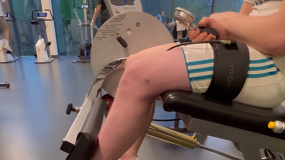
Maintain cardiovascular fitness
- Maintaining cardiovascular fitness is important, especially for runners and athletes. We should focus on what the individual can do. We should see if the individual can use a stationary bike after adjusting the paddles or do aquatic running or other aquatic exercises during this first phase to maintain cardiovascular fitness.
PHASE II: MODERATE LOAD
Before commencing the second phase of ITBS management, make sure that the patient’s pain is within bearable limits, preferably 0-3 on a scale of 10. The pain is subjective and varies considerably from person to person. Just make sure the pain is minimal before starting the closed-chain exercises of the second phase.
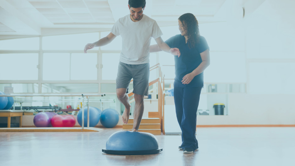
Good hip control
- Restoring good hip and pelvis control is essential to optimally perform closed-chain exercises. We can use visual and verbal cues to correct any deviations from normal while performing closed-chain exercises. We can advise the patient to record themselves or perform the exercise in front of the mirror to have a better understanding of the exercise and incorporate the visual feedback to perform the exercises correctly at home.
Improve tissue capacity
- We will progress in improving the tissue capacity by progressively loading the tissue. Utilizing the principle of progression, we will continually increase the load to allow the tissue to adapt.
Introduce impact drills
- At the end of the second phase, we will introduce impact drills to allow the runners to get ready for the third and last phase of management.
Maintain cardiovascular fitness
- Cardiovascular training should be part of all three phases of ITBS management. Allow the runners to do what they can, like swimming, running in the water, or bicycling using adjustable paddles.
PHASE III: HIGH IMPACT, TOLERANCE, AND READINESS
Before commencing this phase, make sure the patient is pain-free before, during, and after the loading exercises. The patient should be able to walk pain-free for 30 minutes and jog painlessly on the spot for 1 minute. Before progressing to the third phase, the patient’s symptoms should be under control, impact tolerance has improved, and they are now ready to move to the third phase of management.
Improve impact tolerance and restore the plyometric ability
- During the third phase of management, our focus is on the impact tolerance ability of the individual. We must train the ITB to store and release energy like a spring, as other fascial tissues do. We should introduce hopping, skipping, and explosive movements to bring them to the pre-injury level of training.
Gait analysis
- As we have seen that peak hip adduction, medial rotation, and pelvic drop are associated with ITBS, we should observe these while preparing the individual to return to a pre-injury fitness level. We should gradually reduce faulty mechanics because rapidly excluding them may cause other issues in the gait cycle. So progressively excluding these faulty mechanics little by little is the approach we should follow to get optimal results.
WHEN CAN THEY RETURN TO RUNNING?
It might be the most common question the runners will ask. It is important that you guide them through all the phases and explain the importance of returning at a slow and steady pace.
An individual is safe to return to running when they:
- Can jog on the spot for 1 minute?
- Can hop on a single leg for 30 seconds?
- Are ready to run? Do they feel confident about running?
CONSIDERATIONS WHILE RETURNING TO RUNNING
There are certain considerations that should be followed while a patient is returning to running.
- There should be a phased return to running.
- They should consider walking before running and avoid hills initially.
- Start working on frequency and volume before increasing intensity.
REDUCING THE RISK OF RE-INJURY
Reducing the risk of re-injury is an integral part of the overall management of iliotibial band syndrome. We should reflect on three main things when preventing the risk of re-injury.
- After all three phases of management, the patient should return to running gradually. Walk before running, avoid hills initially, and build up training frequency and volume before increasing the intensity.
- No symptoms do not mean that we should stop rehabilitation or management. Most of the time, the runners start the pre-injury level of running without completing the rehabilitation process and end up with re-injury.
- Education of the patient on training errors, training volumes, recovery, and nutrition is as important as the training itself. Without educating the patient, we cannot commence the optimal exercise program and prevent re-injury.
REFERENCES
- Geisler PR. Current Clinical Concepts: Synthesizing the Available Evidence for Improved Clinical Outcomes in Iliotibial Band Impingement Syndrome. J Athl Train. 2021;56(8):805-15.
- KAPLAN EB. The Iliotibial Tract: Clinical and Morphological Significance. JBJS. 1958;40(4):817-32.
- Falvey EC, Clark RA, Franklyn-Miller A, Bryant AL, Briggs C, McCrory PR. Iliotibial band syndrome: an examination of the evidence behind a number of treatment options. Scand J Med Sci Sports. 2010;20(4):580-7.
- Chaudhry H, Schleip R, Ji Z, Bukiet B, Maney M, Findley T. Three-dimensional mathematical model for deformation of human fasciae in manual therapy. J Am Osteopath Assoc. 2008;108(8):379-90.
- Nemeth WC, Sanders BL. The lateral synovial recess of the knee: anatomy and role in chronic Iliotibial band friction syndrome. Arthroscopy. 1996;12(5):574-80.
- Grando H, Chang EY, Chen KC, Chung CB. MR imaging of extrasynovial inflammation and impingement about the knee. Magn Reson Imaging Clin N Am. 2014;22(4):725-41.
- Fredericson M, Cookingham CL, Chaudhari AM, Dowdell BC, Oestreicher N, Sahrmann SA. Hip abductor weakness in distance runners with iliotibial band syndrome. Clin J Sport Med. 2000;10(3):169-75.
- Noehren B, Davis I, Hamill J. ASB clinical biomechanics award winner 2006 prospective study of the biomechanical factors associated with iliotibial band syndrome. Clin Biomech (Bristol, Avon). 2007;22(9):951-6.
- Hamill J, Miller R, Noehren B, Davis I. A prospective study of iliotibial band strain in runners. Clin Biomech (Bristol, Avon). 2008;23(8):1018-25.