ACHILLES TENDINOPATHY; EVIDENCE-BASED ASSESSMENT GUIDELINES, DIAGNOSIS, AND MANAGEMENT PROTOCOLS

DEMOGRAPHICS
Achilles tendinopathy can occur in anyone involved in stretch-shortening cycle activity. In this activity, the Achilles tendon works as an elastic structure like a spring to store and sequentially release the stored energy. In older adults, walking can cause Achilles tendinopathy. In younger populations, it can manifest in athletes like distance runners, track athletes, e.g., sprinters, middle distance runners, and multi-direction sports athletes, like football and hockey players.
ASSESSMENT OF ACHILLES TENDINOPATHY
LOAD TOLERANCE TEST
The load tolerance test allows us to evaluate how much load the Achilles tendon can tolerate. We can do this in two ways:
- We can ask the patient how much load they have with common activities and the pain intensity during these activities.
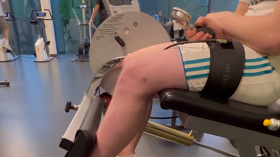
- We can test the load tolerance test in the clinic by manually loading the Achilles tendon. By this, we can objectively assess the capacity of the Achilles tendon and see how much load we can put on the Achilles tendon without provoking the patient’s symptoms.
We should also pay attention to the time it takes for the pain to settle down if it flares during the activities. It can give us essential information about the tolerance of the Achilles tendon.
ACHILLES LOAD TEST
We can load the Achilles tendon by starting with low loads and progressively increasing the loads to see how much load the Achilles can tolerate without aggravating the pain.
We can start loading the Achilles tendon with walking and running and progressing to calf raises and hopping. The image below illustrates the progression of loading the Achilles tendon we can adopt.
 Reproduced from Trustme-ed lectures
ACCEPTABLE PAIN ZONE
While dealing with patients with tendinopathy, we should constrain pain to the acceptable zone. An acceptable zone does not mean the patient should be pain free. People with tendinopathy might have some pain with activities.
 Reproduced from Trustme-ed lectures
According to the numeric pain rating scale, the 0-2 range is the safe zone. The 3-5 range is acceptable, and anything between 6 and 10 is unacceptable and a high-risk zone. Pain up to 5 points is acceptable while loading the tendon.
Another way to state the pain is by using mild, moderate, and severe pain. This approach is usually handy when the patient is unable to assign a number to the intensity of their pain. While using this approach, mild and moderate pain is acceptable, while severe is high-risk and should be avoided.
PHYSICAL FUNCTION CAPACITY OF PATIENTS WITH ACHILLES TENDINOPATHY (1)
Functional capacity assessment of the patients with Achilles tendinopathy can give us useful information about the functional status of the patients. This way, we can assess the errors in the technique and the strength of calf muscles and Achilles tendon. First we will go through some basic concepts regarding the Achilles tendon, and further, we will explain how to assess the physical function capacity of the patients with Achilles tendinopathy.
ENERGY STORAGE TENDON
The Achilles tendon is an energy-storage tendon. The characteristic features of energy storage tendons are:
- These tendons are very elastic and act like a spring
- They have a high injury risk
- They are relatively compliant
- They have a high proportion of glycosaminoglycans
- They have a strain rate of 5-10%
In energy storage tendons, the energy flows from the body to the tendon and again to the body. They store energy and release it when required.
In stretch-shortening cycle activity, strong muscles are essential to harness the stretch in the tendon. If an athlete’s muscles are not strong enough, the energy harness from the energy-storage tendons will be inefficient.
ASSESSMENT OF PHYSICAL FUNCTION CAPACITY
Standing calf raises
- Standing leg raises with the correct technique can give us a good understanding of the physical status of the calf muscles and the Achilles tendon. The tempo for these raises can be 2-3 seconds up and 2-3 seconds down with good plantarflexion and dorsiflexion. We should be cautious about any sway or rollover to the outside of the foot while the patient performs standing calf raises. This way, we can see the endurance by looking at how many repetitions the patients can do without aggravating the pain.
- Hébert et al, in 2017, developed normative values for the standing heel-rise test in healthy adults. They enlisted the estimated number of standing calf raises a healthy individual should be able to do for both the right and left leg and stratified the data according to the individual's age (2).
 Reproduced from Hébert et al, 2017
The normative values are stratified according to the gender and age of the individual. Using these values, we can assess any deficiencies in strength and endurance of the Achilles tendon and the calf muscles. These values can also help us identify any asymmetries between both legs and give us a clues as to where the problem may lie.
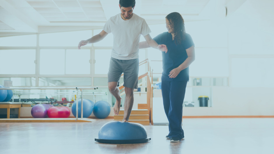
Hopping
- Hopping is a cyclical plyometric activity working on the spring-mass model. During hopping, the ankle joint is the dominant joint utilizing the Achilles tendon as the spring. Hopping can be a useful assessment tool if we look at the height of the hop, ground contact time, and hopping agility. We can also compare the affected leg with the unaffected one and see if there are any asymmetries between both legs. The problems with hopping can be due to pain, calf weakness, capacity issues, or lack of power. This way, hop can help us identify the issues and modify our treatment plan accordingly.
DIAGNOSIS
The diagnosis of Achilles tendinopathy can be simple and complex at the same time. The basic principles for diagnosing Achilles tendinopathy are listed below:
-
Localized pain
-
Onset following a change in load (Walking, running, jumping)
-
Pain aggravated by walking, running, jumping, or any specific activity
-
Proportional load-pain relationship
-
Arising pain/ morning stiffness (Achilles, Plantar, Gluteal)
We need to exclude other possible diagnoses in the ankle region, like the ankle joint, posterior impingement, referred pain, and tibialis posterior.
CLUES TO DIAGNOSING AND POSSIBLE DIAGNOSES FOR THE ACHILLES TENDINOPATHY
The table below enlists the clues to diagnose Achilles tendinopathy and identifying the structures involved.
 Reproduced from Trustme-ed lectures
IMAGING
Ultrasound imaging can be really helpful in case of partial tear of Achilles tendon. Major findings on ultrasound imaging can include:
- Loss of fibrillary structure
- Hypo or anechoic
- Loss of tissue interface border
MANAGEMENT OF THE ACHILLES TENDINOPATHY
EDUCATE THE PATIENT
Patient education plays an essential role in the management of Achilles tendinopathy. Answering the common questions of the patient and giving them a good understanding of the pathology, diagnosis, treatment options, and prognosis can help develop an effective working alliance. It can also help in improving self-efficacy and adherence to the treatment plan (3).
 Reproduced from Trustme-Ed lecture
PAIN MANAGEMENT
Reducing pain is the first step toward the management of Achilles tendinopathy. Before starting any exercise, it is essential to bring the patient's pain down to a minimum. If a patient has a pain intensity of 6 out of 10, managing the pain is more important than starting the isometrics or other strengthening programs. Once the pain is minimum, we can reload the stretch-shortening cycle activity to build strength and endurance of the Achilles tendon.
 Reproduced from Trustme-ed lecture
DIFFERENT APPROACHES TO LOADING PROGRAMS
A systematic review by Malliaras P, Barton CJ, Reeves ND, and Langberg H. compared different Achilles and patellar tendon loading programs to measure the clinical and non-clinical outcomes (3). They compared different approaches, including Alfredson, Stanish and Curvin, Silbernagel, and HSR. The summary of the comparison is illustrated in the image below.
 Reproduced from Malliaras P. et al, 2013
REHABILITATION PHASES FOR ACHILLES TENDINOPATHY
We can divide the Achilles tendinopathy rehabilitation program into four phases starting from the isometric phase and progressing to strength-endurance, maximal strength, and finally to the plyometric.
ISOMETRIC PHASE
At the start, when any isotonic movement is painful, isometrics exercises can make an excellent substitute to build tolerance in the patients and reduce the pain.
Exercises
During the isometric phase, the patient should preferably engage in a home exercise plan and a gym program. A gym program is preferable because we can progress the weight more accurately. The exercise options may include:
- Home options
- The calf wall sits bearing the body weight or half of the body weight
- Standing calf raise hold bearing the body weight or half of the body weight

- Gym options
- Seated calf raise hold bearing the body weight or 1.5 times the body weight
- Leg press holds bearing 1.5 times the body weight

Exercise Prescription
The exercise prescription for the isometric phase can include the following protocols:
- Midrange ankle plantarflexion
- Contraction duration of 10 to 30 seconds
- 5-10 repetitions
- Work to rest ration of 1:1
- Frequency of 1*/day (can be more)
- The intensity of 6-8 out of 10 on the OMNI scale
STRENGTH-ENDURANCE PHASE
After the initial isometric phase, we will progress to some low-load isotonic exercises to build a bit of strength and endurance. We will not be focusing on speed at this stage. We will take things slow and teach the patient the correct technique, including calf raises and endurance calf raises.
We may start with 8-25 repetitions depending upon the patient's functional status. In this phase, we slowly build up strength and endurance.
Exercises
The exercise recommendations for this phase may include the following:
- Standing calf raises bearing the body weight or half of the body weight
- Seated calf raises bearing the body weight or 1.5 times the body weight
 Reproduced from Trustme-ed lecture
Exercise prescription
- Do not go into dorsiflexion if the patient has an insertional problem
- 8-12 repetitions
- 3-4 sets daily
- Work to rest ratio of 1:2
- Frequency of 2-3*/ week
- The intensity of 6-8 out of 10 on the OMNI scale
MAXIMAL STRENGTH PHASE
After building enough strength and endurance during the previous phase, we will progress to the maximum strength phase. In this phase, we will build the capacity to push heavier loads.
Exercise
During the maximal strength phase, we increase the loads to build up tendon and muscle strength. We can progress to isolated eccentric training to build up strength. One way to do it is through calf raises. These may include:
- Standing calf raises bearing body weight or half of the body weight
- Seated calf raises bearing 1.5 times the body weight to 2 times the body weight
Exercise prescription
The exercise prescription may include the following:
- 3-5 repetitions
- 3-4 sets per day
- Work to rest ratio of 1:4
- Frequency of 1-2*/week
- Intensity of 7-9 out of 10 on the OMNI scale
- We will ask the patient for quick concentric contractions to maximize the force generation
STRENGTH-SPEED
In the maximal strength phase, we will try to increase the speed a bit. It does not mean that we are focusing on the speed. Moving with speed will increase the force generation and result in greater strength development.
Exercise
- The sled push with 50% of body weight to 100% of body weight
 Reproduced from Trustme-Ed lecture
The exercise prescription can include the following:
- 4-6 repetitions for each leg
- 3-4 sets per day
- Work to rest ratio of 1:4
- Frequency of 1-2*/ week
- The intensity of 7-9 out of 10 on the OMNI scale
- We will ask the patient for quick concentric contractions to maximize the force generation
SPEED-STRENGTH
In speed-strength training, we will increase the speed at the expense of the load. Increasing the speed would require lowering the loads. So, to get the movement fast, we have to make a force-velocity tradeoff.
PLYOMETRIC PHASE
Plyometric is the last phase of Achilles tendinopathy rehabilitation. Once the patient has developed enough strength and endurance, we can progress to plyometric exercises like hopping.
A study by Sancho et al in 2019, concluded that hopping can be a feasible intervention in recreational runners with Achilles tendinopathy (4).
The variable hopping program from the study is as follows:
Week 1-2
- Low-intensity double leg hop with 2-sets of 20 repetitions once a week with a two-minute rest period between each set
- Medium intensity single leg hop with 2-sets of 20 repetitions once a week with a two-minute rest period between each set
Week 3-4
- Week 1-2 hopping program
- Medium intensity double leg hop with 2-sets of 10 repetitions once a week with a stiff knee and a two-minute rest between each set
- Medium intensity single leg forward and backward hop with-2 sets of 6 repetitions once a week with a two-minute rest period between each set
Week 5-12+
- Week 1-2 hopping program
- Week 3-4 hopping program
- Medium intensity single leg side to-side hop with 2-sets of 6 repetitions once a week with a two-minute rest period between them
- High-intensity single leg hop with 2-sets of 10 repetitions once a week with a stiff knee and a two-minute rest period between them
- High-intensity single leg forward hop with 2-sets of 6 repetitions once a week with a stiff knee and a two-minute rest period between them

References
- Vicenzino B, Vos R-Jd, Alfredson H, Bahr R, Cook JL, Coombes BK, et al. ICON 2019—International Scientific Tendinopathy Symposium Consensus: There are nine core health-related domains for tendinopathy (CORE DOMAINS): Delphi study of healthcare professionals and patients. British Journal of Sports Medicine. 2020;54(8):444-51.
- Hébert-Losier K, Wessman C, Alricsson M, Svantesson U. Updated reliability and normative values for the standing heel-rise test in healthy adults. Physiotherapy. 2017;103(4):446-52.
- Mallows AJ, Debenham JR, Malliaras P, Stace R, Littlewood C. Cognitive and contextual factors to optimise clinical outcomes in tendinopathy. British Journal of Sports Medicine. 2018;52(13):822-3.
- Sancho I, Morrissey D, Willy RW, Barton C, Malliaras P. Education and exercise supplemented by a pain-guided hopping intervention for male recreational runners with midportion Achilles tendinopathy: A single cohort feasibility study. Phys Ther Sport. 2019;40:107-16.